While a good understanding of anatomy is important for the practice of medicine, it's foundational for the practice of surgery. Knowing the relationships between structures can be the difference between curing the patient and causing irreparable harm. Because of its foundational nature, in the U.S. anatomy and physiology is one of the courses taught early in medical school. Around 90% of U.S. medical schools include cadaver dissection as a part of their curriculum.[1] Even as the pedagogy for medical education is transitioning to a flipped classroom model, the importance of in person time studying cadaveric anatomy is not lost on educators. In fact, according to anatomy course directors, one of the most common weaknesses in anatomy curriculum was insufficient dissection time, a problem which was only exacerbated by COVID. [1]
There are many factors that prevent us from maintaining and using an anatomy lab as a part of our medical curriculum here in Burundi. Both the formaldehyde and refrigeration options for preserving cadavers are very expensive. Then comes the practicalities of maintaining constant electrical supply or the safe handling and disposing of large quantities of hazardous chemicals. All this says nothing of the cultural and ethical implications of obtaining cadavers on a regular basis...
So, what are we to do?
Well, 11% of U.S. medical schools also utilize virtual software to enhance, and in some cases replace, the cadaver dissection portion of their anatomy courses. In the post COVID era, a full 23% more plan to incorporate Virtual Reality in their anatomy curricula. [1] While the data is a little old at this point, a 2015 meta-analysis of the educational effectiveness of 3D visualization technologies in teaching anatomy showed that it 1) improved factual knowledge, 2) improved spatial knowledge acquisition, and 3) improved user (aka student) satisfaction as compared to all teaching methods. [2]
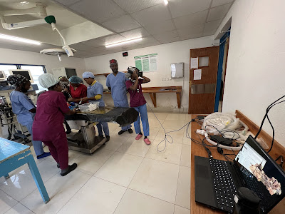
After a few back-and-forth emails, the medical director for The Standford Virtual Heart program graciously provided me with a copy of the software. So after we finished our chapter on congenital heart defects, our residents had a chance to explore the defects and their associated flow patterns and murmurs in virtual reality.
For now, I'm focusing this virtual experience on our current batch of surgical residents. Their need for recalling and understanding anatomy is the most pressing. But the trial run has been well received and quite helpful. I'm excited about the possibility of significantly expanding our use of VR into the anatomy course taught at Hope Africa University.
Afterall, it's hard to build a solid house without a solid foundation...
[1] Shin M, Prasad A, Sabo G, Macnow ASR, Sheth NP, Cross MB, Premkumar A. Anatomy education in US Medical Schools: before, during, and beyond COVID-19. BMC Med Educ. 2022 Feb 16;22(1):103. doi: 10.1186/s12909-022-03177-1. PMID: 35172819; PMCID: PMC8851737.
[2] Yammine K, Violato C. A meta-analysis of the educational effectiveness of three-dimensional visualization technologies in teaching anatomy. Anat Sci Educ. 2015 Nov-Dec;8(6):525-38. doi: 10.1002/ase.1510. Epub 2014 Dec 31. PMID: 25557582.


No comments:
Post a Comment